Animal Cell Electron Microscopy - Light and the Modern Atom : As the name implies, electron microscopes employ an electron beam for imaging.. The animal cell is more fluid or elastic or. Some electron microscopes are capable of magnifying specimens up to 2 million times, while the light microscopes can show a maximum magnification of 2000 times. Generalized cell is used for structure of animal cell and plant cell to present the common parts, appearing in various parts of the bodies of animals and hope you learned a lot about cell structure through our plant cell and animal cell images. These cells may be classified as follows You see that many features are in common.
This tutorial provides a brief description of the subcellular architectures of typical animal and plant cells. Transmission electron microscopy is a proven technique in the field of cell biology and a very useful tool in biomedical research. Generalized cell is used for structure of animal cell and plant cell to present the common parts, appearing in various parts of the bodies of animals and hope you learned a lot about cell structure through our plant cell and animal cell images. Electron microscopy reference describing how backscatter electrons and secondary electrons are detected using scanning electron agrigenomics. Electron microscopy is undoubtedly an indispensable tool in the diagnosis of animal infectious diseases and to investigate the structural analysis of cells and tissues.
Tem Of Animal Cell Photograph by Dr Gopal Murti from images.fineartamerica.com Smooth er plays different functions. Microscopy of wet and untreated whole cells with exceptional ease. Both light microscopes and electron microscopes use radiation (light or electron beams) to form larger and more detailed images of objects than the human eye can produce unaided. • electron microscopes have a much higher resolution than light microscopes. Transmission electron microscopy is a proven technique in the field of cell biology and a very useful tool in biomedical research. These cells may be classified as follows Here, we used high resolution scanning electron microscopy (sem) to study inner cellular structures. Here is an electron micrograph of an animal cell with the labels superimposed:
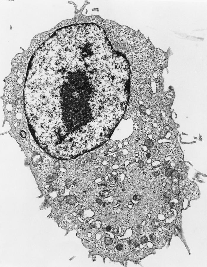
Here is an electron micrograph of an animal cell with the labels superimposed:
Electron microscopy reference describing how backscatter electrons and secondary electrons are detected using scanning electron agrigenomics. Transmission electron microscopy has been an excellent tool, essential for the diagnosis of bacterial and viral animal diseases. Plant, animal and bacterial cells have smaller components each living cells cannot be observed using an electron microscope because samples are placed in a vacuum. You see that many features are in common. Here, we used high resolution scanning electron microscopy (sem) to study inner cellular structures. Electron microscope is a type of microscope that uses electron beam for illumination and creates an enlarged image of the specimen. Smooth er plays different functions. Rank the following biological materials in terms of size, beginning with the smallest (at top) to the largest (at bottom). As the name implies, electron microscopes employ an electron beam for imaging. Microscopy of wet and untreated whole cells with exceptional ease. Innovation and improvements in equipment together with the introduction of new technology have allowed us to improve our knowledge of biological tissues, to visualize. They utilize the same principles behind an optical microscope, but rather than photons or particles of light, concentrate electrons, charged particles located on the outside of. Comparison between a light microscope and an electron microscope:
Smooth er plays different functions. The animal cell is more fluid or elastic or. Light and electron microscopes allow us to see inside cells. Electron microscopes use electron beams some disadvantage of electron microscopes are that they cannot display living specimens in natural colours. This tutorial provides a brief description of the subcellular architectures of typical animal and plant cells.
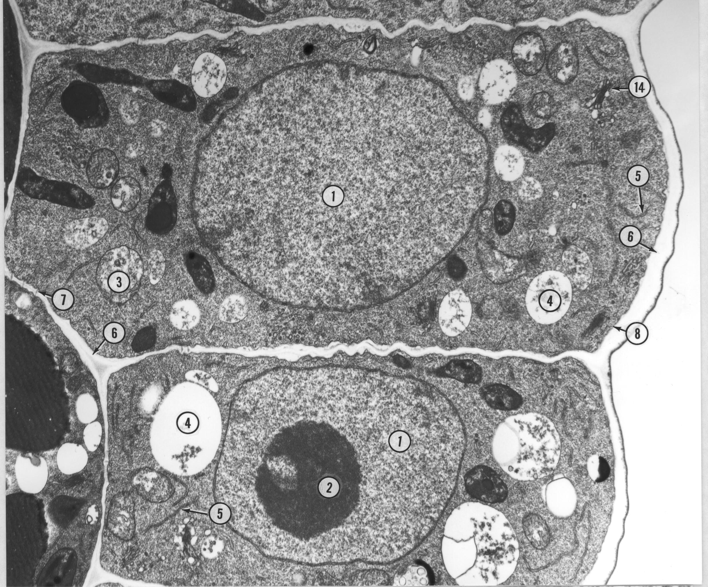
Microscopic Description -- Case 156 | Animal cell ... from i.pinimg.com The smooth endoplasmic reticulum is so named because it appears smooth by electron microscopy. Smooth er plays different functions. Most cells, both animal and plant, range in size between 1 and 100 micrometers and are thus visible only with the aid of a microscope. An electron microscope is a microscope that uses a beam of accelerated electrons as a source of illumination. 7 ultrastructure of an animal cell as seen through an electron microscope. Ultrathin tissue sections are stained with heavy metals (such as osmium tetroxide. Comparison between a light microscope and an electron microscope: Innovation and improvements in equipment together with the introduction of new technology have allowed us to improve our knowledge of biological tissues, to visualize.
The smooth endoplasmic reticulum is so named because it appears smooth by electron microscopy.
The ultrastructure of cells viewed by transmission electron microscopy and scanning electron microscopy. The animal cell is more with light microscopy i can simply scrape some cells from my cheek smear them on a slide and look at them. The animal cell is more fluid or elastic or. Both light microscopes and electron microscopes use radiation (light or electron beams) to form larger and more detailed images of objects than the human eye can produce unaided. During animal cell division, the centrioles replicate (make new copies) and the centrosome divides. A scanning electron microscope (sem) can be used on thicker specimens, such as whole cells or tissues that have been fixed, dried, and coated with a thin metal film. All in all, electron microscopy is not used to see electrons. • electron microscopes have a much higher resolution than light microscopes. Here, we used high resolution scanning electron microscopy (sem) to study inner cellular structures. .gun scanning electron microscopy (fegsem), covering both plant and animal research. Electron microscopy is undoubtedly an indispensable tool in the diagnosis of animal infectious diseases and to investigate the structural analysis of cells and tissues. False plant and animal cells, as well as most microorganisms, can be visualized using brightfield microscopy by enhancing their contrast through staining techniques. 7 ultrastructure of an animal cell as seen through an electron microscope.
During animal cell division, the centrioles replicate (make new copies) and the centrosome divides. Albeit the detail will be minimal without. Transmission electron microscopy is a proven technique in the field of cell biology and a very useful tool in biomedical research. Rank the following biological materials in terms of size, beginning with the smallest (at top) to the largest (at bottom). This tutorial provides a brief description of the subcellular architectures of typical animal and plant cells.
Biology 130 Lab 3 - Electron Micrographs from www4.uwsp.edu Liquid cell electron microscopy is well positioned to explore new frontiers in electrochemistry and catalysis, nanomaterial growth, fluid physics, diffusion, radiation physics, geological and environmental processes involving clays and aerosols, complex biomaterials and polymers, and biological functions. There are two types of electron microscope Share what you learned today with a friend and you'll. A scanning electron microscope (sem) can be used on thicker specimens, such as whole cells or tissues that have been fixed, dried, and coated with a thin metal film. Comparison between a light microscope and an electron microscope: Plant, animal and bacterial cells have smaller components each living cells cannot be observed using an electron microscope because samples are placed in a vacuum. For microscopists on biological applications of field emission gun scanning electron microscopy studies of cells and tissues by both conventional room temperature and cryo electron microscopy. The ultrastructure of cells viewed by transmission electron microscopy and scanning electron microscopy.
Electron microscopy reference describing how backscatter electrons and secondary electrons are detected using scanning electron agrigenomics.
You see that many features are in common. Microscopy of wet and untreated whole cells with exceptional ease. 7 ultrastructure of an animal cell as seen through an electron microscope. Innovation and improvements in equipment together with the introduction of new technology have allowed us to improve our knowledge of biological tissues, to visualize. Ultrathin tissue sections are stained with heavy metals (such as osmium tetroxide. Transmission electron microscopy has been an excellent tool, essential for the diagnosis of bacterial and viral animal diseases. For microscopists on biological applications of field emission gun scanning electron microscopy studies of cells and tissues by both conventional room temperature and cryo electron microscopy. Here, we used high resolution scanning electron microscopy (sem) to study inner cellular structures. This tutorial provides a brief description of the subcellular architectures of typical animal and plant cells. They utilize the same principles behind an optical microscope, but rather than photons or particles of light, concentrate electrons, charged particles located on the outside of. Volume electron microscopy for neuronal circuit reconstruction. Transmission electron microscopy is a proven technique in the field of cell biology and a very useful tool in biomedical research. Here is an electron micrograph of an animal cell with the labels superimposed:


0 Komentar