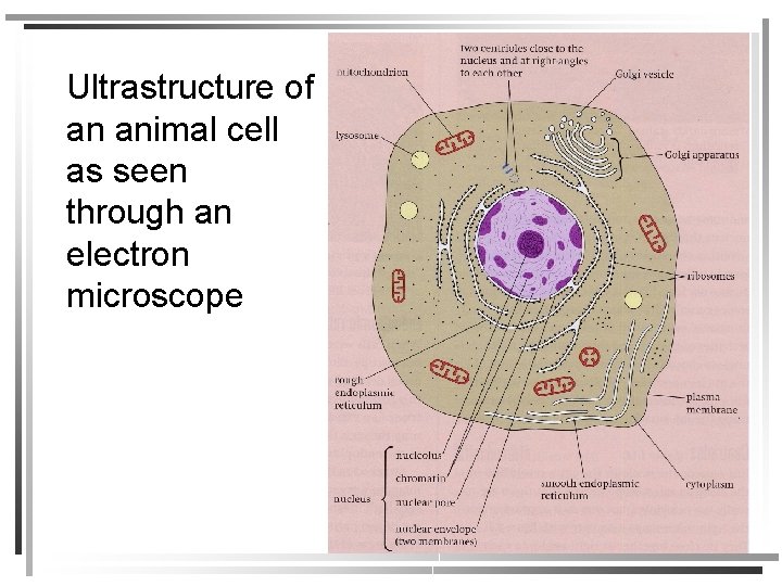
Animal Cell On Microscope / Animal Cells Under Microscope Photos And Premium High Res Pictures Getty Images : 7 ultrastructure of an animal cell as seen through an electron microscope.
Animal Cell On Microscope / Animal Cells Under Microscope Photos And Premium High Res Pictures Getty Images : 7 ultrastructure of an animal cell as seen through an electron microscope.. Onion cells onion cells on low power. Chromatins are of two types, they are euchromatin and heterochromatin. Two slides demonstrating the cell membrane of an animal cell and the cell wall of a plant cell. Find the perfect animal cell microscope stock photos and editorial news pictures from getty images. Digital artwork creative graphic design.
Plant cells have cell walls, one large vacuole per cell, and chloroplasts, while animal cells will have a cell membrane only. We zoom in on an individual cell at 28:00 we look at the cheek cells. Use sunlight to make food through photosynthesis. Generalized cell is used for structure of animal cell and plant cell. Cell division under the microscope isolated on a white with alpha map.
Animal And Plant Cells Microscope Slide Set Microscope Sample Slides Amazon Com Industrial Scientific from m.media-amazon.com • electron microscopes have a much higher resolution than light microscopes. Below the basic structure is shown in the same animal cell, on the left viewed with the light microscope, and on the right with the transmission electron microscope. Plant and animal cell, microscopes. A specimen is a small part or slice, or an. Review comparing animal and plant cell lab. Animal cell under a light microscope. All animal and plant cells are enclosed or surrounded by a cell membrane as we learned before. Find the perfect animal cell microscope stock photos and editorial news pictures from getty images.
Students know how prokaryotic cells, eukaryotic cells (including those from plants and animals), and viruses differ in complexity and general structure.
Cell parts that can be observed using a compound light. A cell carries out all the processes of the body which includes producing energy and storing it when seen under the microscope, the chromatin will have an appearance like beads on a string. Cell division under the microscope isolated on a white with alpha map. A cell is a very tiny structure which exists in living bodies. All animals are composed of cells, a principle that was elucidated by theodor schwann in 1839. Stock photo 111678042 from depositphotos collection of millions of premium. Plant cell has cell wall and cell membrane and animal cell has vacuole and nucleus. Unlike animal cells (such as cheek cells) the cell wall of an onion and other plants are made up of cellulose, which protects the cell and maintains its shape. They have a distinct nucleus with all cellular organelles under the microscope, an animal cell shows many different parts called organelles, that work together to keep the cell functional. We zoom in on an individual cell at 28:00 we look at the cheek cells. Most of the cells that compose animal tissues and organs however, with the aid of a microscope, one can observe the basic animal cell structure. 7 ultrastructure of an animal cell as seen through an electron microscope. Electron microscopes use electron beams focused by electromagnets to magnify and resolve microscopic specimens.
They have a distinct nucleus with all cellular organelles under the microscope, an animal cell shows many different parts called organelles, that work together to keep the cell functional. Examining animal cells under the microscope. 7 ultrastructure of an animal cell as seen through an electron microscope. Prepared microscope slides set for students basic biological science education, 25pcs animal plant insect bacteria specimens, containing wooden. Human or animal cells on blue background.
What Are The Visible Plant Animal Cell Organs On Light Microscope Quora from qph.fs.quoracdn.net Move your slide so that your field of view is centered on. We zoom in on an individual cell at 28:00 we look at the cheek cells. What cell organelles can be seen under the electron microscope but not with the light microscope and their functions in the cell? Set up your microscope, place the onion root slide on the stage and focus on low (40x) power. All animal and plant cells are enclosed or surrounded by a cell membrane as we learned before. Select from premium animal cell microscope of the highest quality. Actually the cells in your mouth can be taken out easily though it will be painful to take any cell out. Cheek cells under the microscope.
Two slides demonstrating the cell membrane of an animal cell and the cell wall of a plant cell.
We zoom in on an individual cell at 28:00 we look at the cheek cells. Digital artwork creative graphic design. 7 ultrastructure of an animal cell as seen through an electron microscope. Cell parts that can be observed using a compound light. • electron microscopes have a much higher resolution than light microscopes. Huge collection, amazing choice, 100+ million high quality, affordable rf and rm images. A cell carries out all the processes of the body which includes producing energy and storing it when seen under the microscope, the chromatin will have an appearance like beads on a string. Set up your microscope, place the onion root slide on the stage and focus on low (40x) power. All animal and plant cells are enclosed or surrounded by a cell membrane as we learned before. Below the basic structure is shown in the same animal cell, on the left viewed with the light microscope, and on the right with the transmission electron microscope. Electron microscopes use electron beams focused by electromagnets to magnify and resolve microscopic specimens. Even more amazing is to see your own cells under the microscope. All animals are composed of cells, a principle that was elucidated by theodor schwann in 1839.
Plant and animal cell, microscopes. Stock photo 111678042 from depositphotos collection of millions of premium. Animal cells are of various sizes and have irregular shapes. Review comparing animal and plant cell lab. Human or animal cells on blue background.
Structure Of Plant And Animal Cells Under An from slidetodoc.com Chromatins are of two types, they are euchromatin and heterochromatin. Established lines and primary cultures of human and animal cells can be extremely. Most of the cells size range between 1 and 100 micrometers and are visible only with the microscope. Electron microscopes use electron beams focused by electromagnets to magnify and resolve microscopic specimens. Select from premium animal cell microscope of the highest quality. Huge collection, amazing choice, 100+ million high quality, affordable rf and rm images. Cell division under the microscope isolated on a white with alpha map. Animal cells are of various sizes and have irregular shapes.
Most of the cells that compose animal tissues and organs however, with the aid of a microscope, one can observe the basic animal cell structure.
Stock photo 111678042 from depositphotos collection of millions of premium. A cell is a very tiny structure which exists in living bodies. Review comparing animal and plant cell lab. Two slides demonstrating the cell membrane of an animal cell and the cell wall of a plant cell. Cheek cells under the microscope. Actually the cells in your mouth can be taken out easily though it will be painful to take any cell out. Microscope artificial cell synthesis animal human designer cell biochemistry. • electron microscopes have a much higher resolution than light microscopes. Microscopes produce magnified images of cells so we can study them in detail. We cannot even say the single animal cell perform which function.but as the animal cell is composed up of several organelles and these organelles perform. Even more amazing is to see your own cells under the microscope. However, as you probably noticed in the previous to view cells under a microscope, we need to make and prepare something called a specimen on a slide. Most of the cells size range between 1 and 100 micrometers and are visible only with the microscope.


0 Komentar